Cell Health & Metabolism Solutions for Cell Therapy
Monitoring cell health and metabolism is crucial in cell therapy as they directly impact the efficacy, safety, and overall therapeutic potential of the final cell products. Our diverse portfolio of sensitive, add-and-read assays can help you answer questions about the viability, proliferation and metabolism of your cells.
Learn how T cell activation protocols are optimized to enrich stem cell memory phenotypes in this poster.
Are My Cells Viable?
CellTiter-Glo® Luminescent Assays measure cell viability based on ATP quantitation, which indicates metabolically active cells. These assays are run in a homogeneous ‘add-mix-measure’ format allowing for a fast and easy workflow, especially when integrated with the MyGlo™ Reagent Reader or GloMax® Microplate Readers. These assays are used to assess cell viability in multiple model systems and sample types, including monolayer cell cultures, 3D microtissues, organoids, primary cells and stem cells.

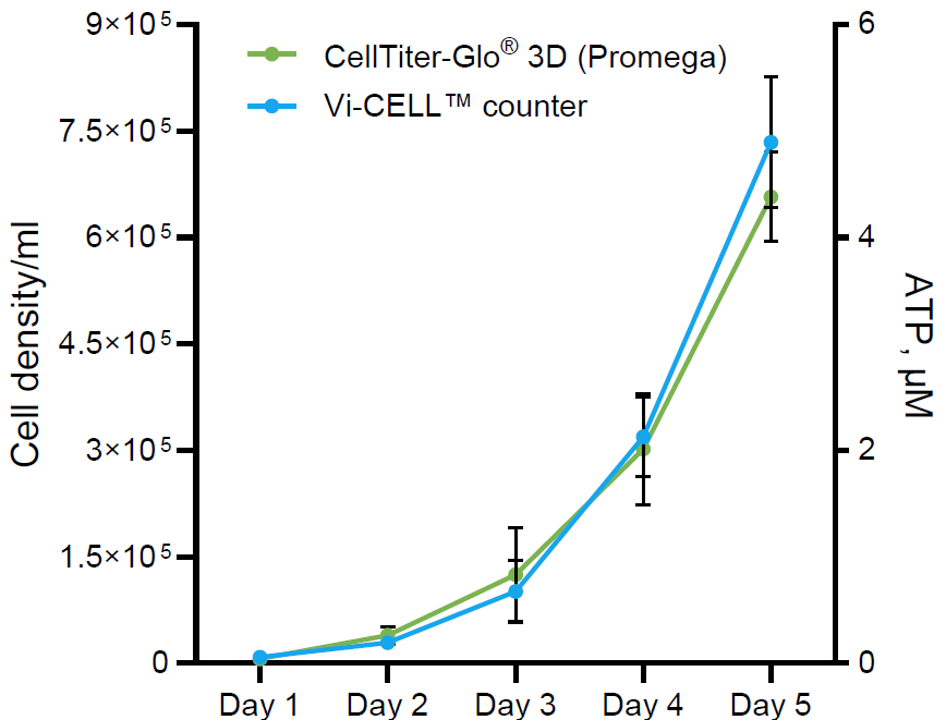
The quantification of viable Human Adipose Derived Stem Cells (hADSCs) during a 5 day-expansion in the BioBLU® 0.3c Single-Use Bioreactor using CellTiter-Glo® 3D Cell Viability Assay in comparison to cell counts from Vi-CELL™ automated cell counting device. Data represents the average of six bioreactors (n=6).
See selected citations on how our cell viability assays are used in cell therapy development:
| Product Used | Citation |
|---|---|
| CellTiter-Glo® 2.0 Cell Viability Assay was used to measure the killing of CD20-expressing cancer cells by a novel allogeneic Rituximab-conjugated γδ T cells. | Li, H.K. et al. (2023) A novel allogeneic Rituximab-conjugated gamma delta T cell therapy for the treatment of relapsed/refractory B-cell lymphoma. Cancers 15, 4844. |
| CellTiter-Glo® 2.0 Cell Viability Assay was used to assess the ablation of iPSCs modified and induced to express a suicide gene in vitro. | Dahlke, J. et al. (2021) Efficient genetic safety switches for future application of iPSC-derived cell transplants. J. Pers. Med. 11, 565. |
| CellTiter-Glo® Luminescent Cell Viability Assay was used to measure target tumor cell killing by CAR-T cells in vitro. | Kuo, Y.-C. et al. (2022) Antibody-based redirection of universal Fabrack-CAR T cells selectively kill antigen bearing tumor cells. J. Immunother. Cancer 10, e003752. |
| LDH-Glo™ Cytotoxicity Assay was used to measure the in vitro cytotoxicity of CAR-T cells redirected to CD30 on peripheral T-cell lymphoma cells. | Wu, Y. et al. (2022) A new immunotherapy strategy targeted CD30 in peripheral T-cell lymphomas: CAR-modified T-cell therapy based on CD30 mAb. Cancer Gene Ther. 29, 167-177. |
Are My Cells Proliferating?
The easy, add-mix-measure plate-based Lumit® hKi-67 Immunoassay uses the common marker Ki-67 to measure cell proliferation. The assay overcomes challenges associated with other common techniques such as cumbersome wash steps, metabolic activity misrepresenting proliferation, and the need for specialized equipment. It can help you reduce labor and prep time, make quicker decisions, and increase confidence in your data.


Human CD8+ T cells (StemCell Tech) plated at 80,000 cells per well were treated with CD3/CD28 T cell activator with (orange) and without (blue) 10ng/ml IL-2 for 48 hours. Upregulation of Ki-67 is observed with the Lumit® hKi-67 Immunoassay before T cell proliferation (which begins >72 hours after activation), demonstrating use of this assay as an early indicator of proliferation.
Are My Cells Metabolically Fit?
Cell metabolism is crucial in cell therapy development and manufacturing because it influences cell growth, differentiation, and function, which are essential for producing high-quality, therapeutic cells. Understanding and optimizing metabolic pathways can enhance cell yield, viability, and therapeutic efficacy, ensuring the success and scalability of cell therapies. We offer a diverse portfolio of sensitive, add-and-read bioluminescent assays to measure metabolic activity, including glucose uptake, lactate, glutamine, oxidative stress and NAD(P)H.
Cell metabolic state can be evaluated by:
- Monitoring changes in metabolites in cell culture medium
- Profiling intracellular key metabolic co-factors (NAD(P)/NAD(P)H and metabolites
- Measuring activity of dehydrogenases, key regulators of major metabolic pathways

Monitoring Metabolism in T Cells
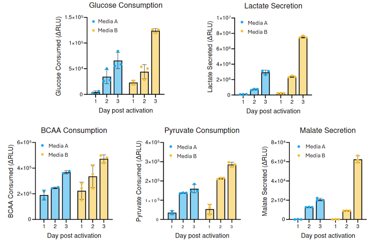
T cells with a stem cell memory phenotype (Tscm) are known for their prolonged in vivo presence and function compared to other types. The generation of Tscm cells holds significant promise for immunotherapies, such as CAR-T. Optimizing and monitoring culture conditions using cell health and metabolism assays can help identify predictive markers of Tscm enrichment, yielding more desired Tscm cells. In addition, metabolic measurements can serve as early predictors of T-cell phenotypic profiles.


Metabolic profiles of activated T cells vary depending on culture conditions. See more data in this poster: Optimization of T Cell Activation Protocols to Enrich Stem Cell Memory Phenotypes
Learn more about monitoring metabolism in T cells:
Poster
Optimization of T Cell Activation Protocols to Enrich Stem Cell Memory Phenotypes
Learn how metabolic measurements can be used as an early screen for predicting phenotypic profiles of expanded T-cells.
Publication
Metabolic Priming for Manufacturing CAR-T Cells
Learn how our metabolite detection assays were used to optimize media and growth conditions during CAR-T cell manufacturing to enhance persistence, potency and memory formation.
Poster
Simple Cell-Based Assays for Profiling Metabolism in Cell Therapy Products
Learn how our bioluminescent metabolite detection assays can be used to profile metabolic changes during T cell activation.
Monitoring Metabolism for hMSC Expansion
The success of human mesenchymal stem cell (hMSC) therapy depends on expanding metabolically fit cells suitable for in vivo function. Monitoring nutrients such as glucose consumption and lactate production during expansion, oxidative phosphorylation, and intracellular glutamine and glutamate levels is important for consistent and uniform cell expansion.

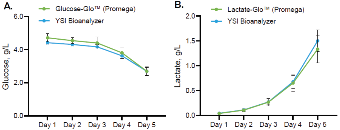
Glucose consumption (Panel A) and lactate secretion profile (Panel B) of hADSC cultured for 5 days in the BioBLU® 0.3c Single-Use Bioreactor controlled by a DASbox® Mini Bioreactor System. Data represents the average of six bioreactors (n=6) and shows Glucose-Glo™ and Lactate-Glo™ Assays generated the same profiles as those from YSI Bioanalyzer.

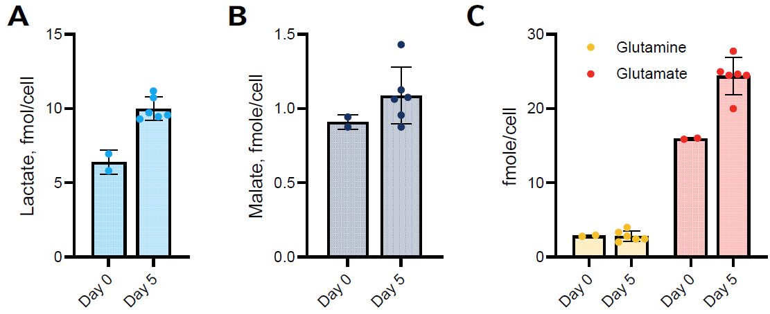
Intracellular metabolite levels. Cells were removed from microcarriers on day 5 and compared to day 0. A total of 3,000 cells/well were assayed in a 384-well low volume plate. Lactate (Panel A), malate (Panel B), and glutamine/glutamate (Panel C) were assayed using metabolite detection assays. Data represents the average of six bioreactors (n=6).
Download this app note to see more data: Metabolic phenotype preservation and suitability of human mesenchymal stem cells cultivated in stirred tank bioreactors for cell therapy applications.