Fluorescence Microscopy
Learn the basics of fluorescence microscopy, including its applications and advantages.
Need ligands with improved brightness, quantum yield, and longer fluorescence lifetime?
What is Fluorescence Microscopy?
Fluorescence microscopy is a core imaging technique that exploits the principles of fluorescence to visualize and study the properties of organic and inorganic substances, particularly biological specimens. By tagging specific molecules with fluorescent probes, researchers can observe the localization, distribution, and dynamics of these molecules within complex biological systems.
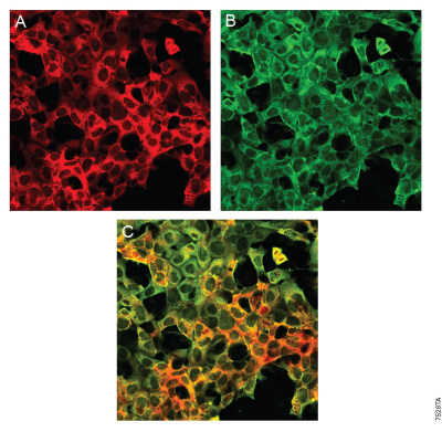
Distinct labeling of a single HaloTag® 7 fusion by a HaloTag® Ligand and Anti-HaloTag® pAb. See details.
How Does Fluorescence Microscopy Work?
The basic principle of fluorescence microscopy involves the excitation of fluorophores by a specific wavelength of light. Upon excitation, these fluorophores emit light at a longer wavelength, which is then detected to produce an image. The difference between the excitation and emission wavelengths is known as the Stokes shift, and it allows for the separation of emitted fluorescence from the excitation light using optical filters.

Janelia Fluor® HaloTag® Ligands: Brighter Ligands, No-Wash Protocol
Unlock new possibilities in fluorescence imaging with photoactivatable Janelia Fluor® Ligands designed to deliver superior brightness, higher quantum yield, and extended fluorescence lifetimes. Explore both membrane-permeable and impermeable options.
The HaloTag® platform enables the use of multiple Janelia Fluor® Ligands with a single construct. Whether working on multiplex experiments or leveraging instruments with different filter sets, this platform ensures maximum adaptability.

What is Fluorescence Microscopy Used For?
Fluorescence microscopy has a wide range of applications in various scientific fields. A selection of these applications are highlighted below:
Molecular Biology
Gene expression can be investigated through tagging mRNA or specific protein and tracking both synthesis, trafficking, and degradation in live cells. This enables visualization of protein-protein interactions, membrane protein localization and signaling pathways in response to environmental changes or stimuli, such as growth factors, or stress.
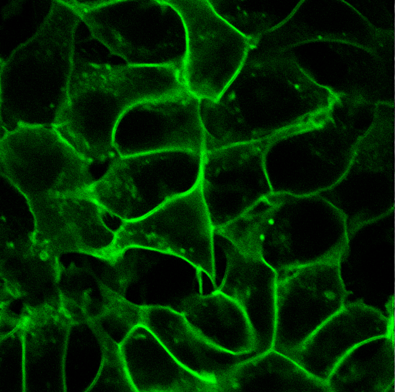
Live-cell labeling of cell surface HaloTag® fusion proteins.
Cell Biology
Fluorescence microscopy enables visualization of cellular structures, such as organelles, cell membranes, and cytoskeletal components. Localization of proteins can also be tracked in cells, providing insight into their cellular function. You can also visualize distinct protein dynamics, such as their change in shape, how they interact with other cellular components over time, and general movement.
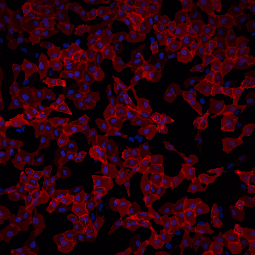
Cell-impermeable ligand imaging using Janelia Fluor® HaloTag® Ligands.
Neuroscience
Mapping neural circuits, including neuronal connections to reveal signal transmission. Specific synaptic activity can also be observed through tracking of individual synapses. Neurotransmitter dynamics can also be observed, such as release, diffusion, and reuptake.
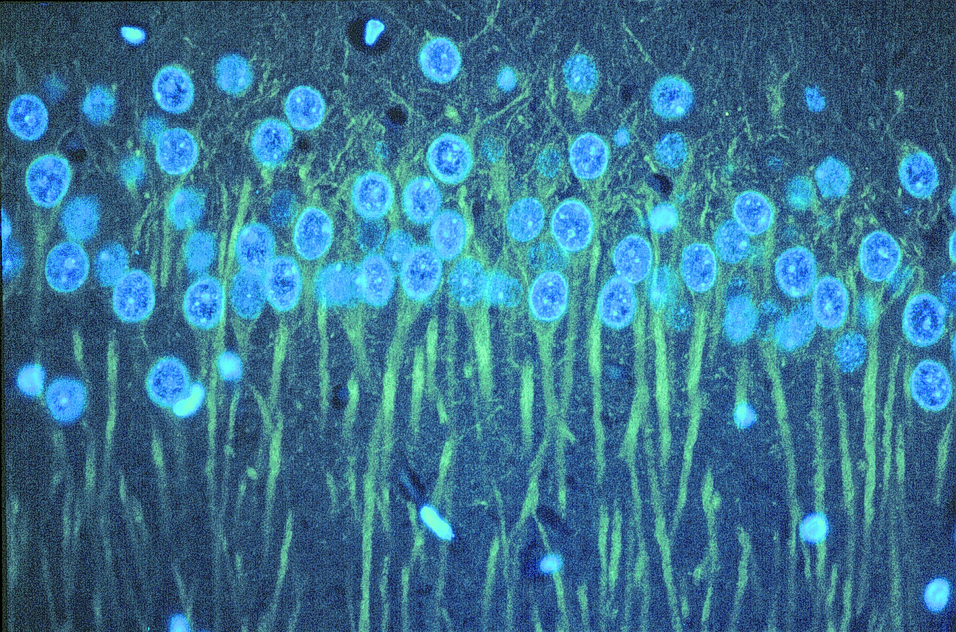
Immunostaining for βIII tubulin in rat cerebellum using Anti-βIII Tubulin mAb.
Webinar: Using HaloTag® Technology and Janelia Fluor® Dyes for Live Cell Single-Molecule Imaging
Explore how single-molecule imaging reveals sub-cellular dynamics and biochemical properties of biomolecules in their native context. In this webinar, we discuss how HaloTag® Technology and Janelia Fluor® Ligands enable precise single-molecule detection, uncovering molecular heterogeneity and challenging biological paradigms.
Fluorescence Microscopy Advantages and Disadvantages
Advantages
Targeted Labeling
Fluorescence microscopy allows for the specific labeling of structures, molecules, or proteins using fluorescent dyes or tags. This specificity enables researchers to highlight components within complex biological samples.
Multiplexing
Multiple fluorescent labels can be used simultaneously, allowing for the visualization of several targets within the same sample.
High Contrast
Fluorescence microscopy provides high-contrast images, as the background is typically dark, and only the fluorescently labeled structures are visible. This enhances the visibility of specific features.
Wide Range of Applications
Fluorescence microscopy is versatile and can be applied to a broad range of samples, including fixed and live cells, tissues, and whole organisms.
Adaptability
Various fluorescent probes and techniques (e.g., FRET, FRAP, STORM) can be adapted to study diverse biological processes and interactions.
Disadvantages
Autofluorescence
Background fluorescence can make it difficult to determine where your fluorescent tag is located. Autofluorescence can arise from background proteins or the sample plate/slide.
Photobleaching
Although the sensitivity of fluorescent proteins to light can vary widely, there is typically a reduction in light emission over time. This means that your fluorescent tags will become more difficult to discern over long imaging sessions.
Want to avoid problems such as autofluorescence and photobleaching? This is possible with bioluminescence imaging. Learn more about bioluminescence imaging in cells and whole animals.
Using HaloTag® Technology to Image Advanced Cell Models
Learn how Janelia Fluor® Ligands are used to image advanced cell models (e.g., spheroids).
How to Study Protein Localization, Trafficking and Turnover
See how fluorescence microscopy with HaloTag® Technology can support your research.
Webinar: Tracking Cellular Protein Localization and Movement in Cells
Learn how HaloTag® Technology works and its applications in protein imaging.
Other Imaging Resources
Imaging
Learn about various imaging techniques, including live cell imaging, in vivo imaging, bioluminescence imaging and fluorescence microscopy.
Live Cell Imaging
Learn about live cell imaging techniques, advantages and applications.
Bioluminescence Imaging
Learn about bioluminescence imaging in whole animals and in live cells.