Imaging Devices
The GloMax® Galaxy Bioluminescence Imager is an easy-to-use, affordable imaging device with bioluminescence, fluorescence and brightfield capabilities. Designed for low-throughout assay validation, the GloMax® Galaxy Bioluminescence Imager is designed for the imaging of NanoLuc® Luciferase technologies, such as HiBiT, NanoBiT and NanoBRET.
Imaging Devices Products
GloMax® Galaxy Bioluminescence Imager System
A bioluminescence microscope that enables functional imaging of NanoLuc® Technologies.
GloMax® Galaxy Bioluminescence Imager Accessories
Accessories include the Tokai Environment Chamber, fluorescence modules and imaging adaptors.
GloMax® Galaxy Bioluminescence Imager Service and Support Products
Covered by 1-year Standard Warranty upon purchase.

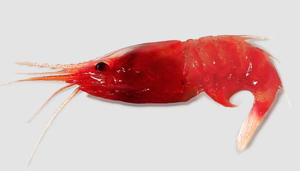
Engineering NanoLuc® Luciferase from a Deep-Sea Shrimp
Read this paper to learn how NanoLuc® Luciferase was developed by merging optimization of protein structure and a novel substrate.

Video: Exploring a Segmentation Clock Model Using Bioluminescent Live-Cell Imaging
This team used the Nano-Glo® Live-Cell Assay System to study temporal patterns in cellular development.

Video: HiBiT Protein Tagging System
Watch this video to learn how HiBiT can solve your protein tagging problems and more.
What Does Bioluminescence Imaging Enable?
When performing plate-based assays, it can often be challenging to determine whether the signal obtained is representative of a significant portion of the cell population. Cellular imaging enables one to see inside a cell, to visualize and analyze the processes occurring within a living cell. Cellular Bioluminescence Imaging (BLI) leverages genetically modified cells that express bioluminescent proteins, allowing high-resolution, real-time monitoring of dynamic cellular and subcellular events. Unlike fluorescence imaging, BLI does not require external light for stimulation. Instead, an enzymatic substrate is introduced to produce a luminescent signal. This method reduces the risks of phototoxicity and photobleaching, which are common concerns in fluorescence imaging.
The brightness and specificity of bioluminescent reporters make them ideal for various research applications. In the study of genetic expression and regulation, they allow researchers to link luciferase reporters to specific gene promoters, observing the activation or suppression of genes in response to different stimuli, drugs or environmental conditions. For studying protein-protein interactions, techniques like bioluminescence resonance energy transfer (BRET) enable the detection of fluorescent signals without the need of external light stimulation. By tagging signaling molecules with HiBiT, researchers can visualize and quantify the activation of specific cell signaling pathways in response to various stimuli without impeding endogenous cellular activity. It can also be used to monitor viral and bacterial infections, with pathogens engineered to express bioluminescent reporters allowing for real-time tracking of infection processes, replication rates, and the effects of antimicrobial treatments.

