Selective Cytochrome P450 3A7 Enzyme Assay Using a Novel Bioluminescent Probe Substrate
Promega Corporation
Abstract
This is part of a series of seven articles describing new bioluminescent substrates to be used for drug screening and assaying enzymatic activity.
Introduction
CYP3A enzymes are cytochromes P450 (CYPs) that oxidize many endogenous compounds, drugs and other xenobiotics (1) . CYP3A7 is the predominant CYP3A enzyme in fetal and neonatal liver. CYP3A7 expression declines after birth in parallel with a dramatic increase in CYP3A4 and CYP3A5, which become the most abundant CYPs in adult liver and intestine (2) . CYP3A7 assay chemistries are frequently cross-reactive with CYP3A4 and CYP3A5 and commonly require chromatographic steps that limit the assays in terms of ease-of-use and throughput. Consequently, there is a need for a simple, rapid multiwell plate-based CYP3A7 assay that is met by the luminogenic assay described here.
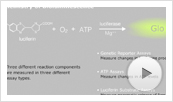
Luminogenic CYP assays use prosubstrates for the light-generating reaction of firefly luciferase. CYPs convert the prosubstrates to luciferin, which makes light in a second reaction with luciferase (3) . The amount of light generated is proportional to the amount of luciferin produced by the CYP and therefore to CYP enzyme activity (4) . Multiple CYP enzymes are encoded by families of genes in humans and other organisms (5) . The CYP enzyme selectivity for a given luminogenic substrate depends on the nature of the derivatization on the luciferin structure.
Here we demonstrate that 2,2′-(6,6′-(1,3-phenylenebis(methylene))bis(oxy)bis(benzo[d]thiazole-6,2-diyl))bis(4,5-dihydrothiazole-4-carboxylic acid), a bis-luciferin referred to herein as Luciferin-3A7, is a luciferase prosubstrate that is selectively converted to luciferin by the human CYP3A7 enzyme with little or no detectable activity with CYP3A4 or CYP3A5. We describe how this luciferase prosubstrate is used in a luminogenic CYP3A7 biochemical assay to detect CYP3A7 activity and CYP3A7 inhibitors.
Materials and Methods
The CYP3A7 enzyme assay was performed using instructions in the P450-Glo™ Assay Technical Bulletin #TB325 and P450-Glo™ Screening Systems Technical Bulletin #TB340. The CYP enzymes used were recombinant human forms in microsomes from insect cells that coexpress a human CYP cDNA with P450 reductase, or P450 reductase plus cytochrome b5 (Gentest™ Supersomes™, BD Biosciences). A 5mM stock solution of Luciferin-3A7 substrate (molecular weight = 662.77) was prepared in 100mM KPO4 buffer. The 50µl CYP3A7 enzyme assays were performed using 30µM Luciferin-3A7 substrate or as indicated in figure legends, 100mM KPO4 (pH 7.4), 20nM CYP3A7 enzyme coexpressed with P450 reductase and cytochrome b5 (1pmol/50µl reaction), and 1X NADPH Regeneration System (Cat.# V9510).
Assays were assembled and performed in opaque white 96-well plates (e.g., white polystyrene, 96-well plates [Costar Cat.# 3912]). After incubating reactions for 20 or 30 minutes at 37°C or room temperature (20–23°C), 50µl of Luciferin Detection Reagent (LDR; Cat.# V8920, V8921) was added to each 50µl CYP reaction to stop the reactions and initiate luminescence. Luminescence was read after 20 minutes using the GloMax® 96 Microplate Luminometer (Cat.# E6501) and reported in relative light units (RLU).
For convenience, you can prepare a 4X concentrated enzyme/buffer/substrate mix (400mM KPO4 [pH 7.4], 120µM Luciferin-3A7, 80nM CYP3A7), 4X concentrated test compound solution (e.g., for CYP inhibition assays) and 2X concentrated NADPH Regeneration System. For a 50µl reaction, combine 12.5µl of the 4X enzyme mixture with 12.5µl of 4X test compound and initiate the reaction by adding 25µl of the NADPH Regeneration System (6) , (7) .
Results and Discussion
Substrate Selectivity
The putative luminogenic CYP substrate, Luciferin-3A7, was initially screened for activity in the luminescent assay format against 21 recombinant human CYP enzymes (Figure 1). Under the conditions used in Figure 1, we observed minor reactivity with CYP2C8 and CYP3A4, but the most prominent activity was with CYP3A7.
 Figure 1. CYP enzyme selectivity for the Luciferin-3A7 substrate.
An anticipated reaction scheme with Luciferin-3A7 (I) is shown at the top. Intensity of luminescence generated by LDR depends on the conversion of I to luciferin (II). Fifty microliter reactions with 5µM Luciferin-3A7 and 20nM recombinant human CYP enzymes in 96-well plates were incubated for 30 minutes at 37°C. Values are mean ± SD, n = 3.
Time Dependence
At both room temperature and 37°C, a time-dependent linear increase in luminescence was observed up to 30 minutes (Figure 2). The modest deviations from linearity after 30 minutes may reflect instability of the CYP3A7 enzyme.
 Figure 2. Time course of the CYP3A7 reaction with Luciferin-3A7.
Time-dependent changes in net luminescence were monitored at room temperature (22°C) and 37°C. Fifty microliter reactions were performed in 96-well plates with 50µM Luciferin-3A7 and 20nM CYP3A7 in 100mM KPO4 (pH 7.4). CYP reactions were initiated by staggered addition of the NADPH Regeneration System. All CYP reactions were simultaneously terminated and luciferase reactions initiated by adding 50µl of LDR. Zero-time values were measured in samples where the NADPH Regeneration System was withheld until after LDR addition. The luminescence shown was total luminescence of samples minus luminescence of control samples with no CYP enzyme. Values are mean ± SD, n = 3.
Substrate Concentration
A concentration-dependent increase in CYP3A7 activity was observed up to a maximal rate between 50–100µM of Luciferin-3A7, with a slight decrease at 200µM (Figure 3). The data fit well to a substrate inhibition model (R2=0.99) described by the equation Y = (Vmax × S)/(Km + S + [S × S/Ki]), where S = substrate concentration, Y = activity in relative light units (RLU), Km = the half maximal rate, and Ki = the substrate inhibition constant. The calculated Km and Ki values are 33µM and 189µM, respectively.
 Figure 3. Km measurement of CYP3A7 using Luciferin-3A7.
Reactions with 20nM CYP3A7 and 100mM KPO4 (pH 7.4) were incubated for 20 minutes at 37°C. The curve fit to a substrate inhibition model was performed using GraphPad Prism® software. The luminescence shown (mean ± SD, n = 3) is total luminescence of samples minus luminescence of control samples lacking CYP enzyme at each substrate concentration.
Enzyme Concentration
Signals from a reaction at 30µM of Luciferin-3A7 increased in a linear fashion with increasing CYP3A7 enzyme concentration up to 10nM of enzyme (Figure 4). The lowest concentration of CYP3A7 tested (0.625pM) gave activity above the limit of detection (LOD). By extrapolation of a linear regression for values ≤10nM of CYP3A7 enzyme, the LOD for these reaction conditions is 0.3nM of CYP3A7, when LOD is defined as the mean of the background plus three standard deviations.
 Figure 4. Titration of the CYP3A7 enzyme using Luciferin-3A7.
Reactions were performed with a range of CYP3A7 concentrations. Reactions used 30µM Luciferin-3A7 and were incubated for 30 minutes at 37°C. The luminescence shown (mean ± SD, n = 3) is total luminescence of samples minus luminescence of control samples with no CYP enzyme.
CYP3A7 Inhibition
We tested CYP3A7 inhibition using the CYP3A7 assay with 30µM of Luciferin-3A7, the approximate Km concentration (Figure 5). Dose-dependent inhibition by ketoconazole was observed with an IC50 of 0.4µM, which correlates well with a previously published value for CYP3A7 inhibition by ketoconazole (8) .
 Figure 5. Measuring CYP3A7 inhibition using Luciferin-3A7.
Using 20nM CYP3A7, enzyme inhibition was assayed using 30µM Luciferin-3A7 in the presence of ketoconazole at the indicated concentrations. The ketoconazole was diluted from a 25mM stock solution in DMSO, and the contribution of this vehicle was kept constant at 0.1% in all reactions. Reactions were incubated at 37°C for 20 minutes before adding LDR. Background luminescence from control samples with no CYP enzyme (mean background = 22,573 RLU) was subtracted to give the net luminescence values shown (mean ± SD, n = 3).
Conclusion
Luciferin-3A7, a novel luciferin derivative, is a luminogenic cytochrome P450 substrate with excellent selectivity for the human CYP3A7 enzyme. The application of Luciferin-3A7 to the luminogenic CYP enzyme assay approach harnesses the exquisite sensitivity, selectivity and simplicity of bioluminescence. This provides for an easy, rapid, multiwell plate-based CYP3A7 enzyme assay that can be used to screen for potential CYP3A7 inhibitory compounds.
Related Products
 Learn about the chemistry of the bioluminescent reaction and its use in a broad range of assays.
Related Resources
Article References
- Hines, R.N. and McCarver, D.G. (2002) The ontogeny of human drug-metabolizing enzymes: Phase I oxidative enzymes. J. Pharm. Exp. Ther. 300, 355–60.
- Stevens, J.C. (2006) New perspectives on the impact of cytochrome P450 3A expression for pediatric pharmacology. Drug Disc. Today 11, 440–5.
- Branchini, B.R. et al. (1998) Site-directed mutagenesis of histidine 245 in firefly luciferase: A proposed model of the active site. Biochemistry 37, 15311–9.
- Cali, J.J. et al. (2006) Luminogenic cytochrome P450 assays. Exp. Op. Drug Metab. Toxicol. 2, 629–45.
- Guengerich, F.P. (2006) Cytochrome P450s and other enzymes in drug metabolism and toxicity. AAPS J. 8, E101–11.
- P450-Glo™Assays Technical Bulletin, TB325, Promega Corporation.
- P450-Glo™ Screening Systems Technical Bulletin, TB340, Promega Corporation.
- Stresser, D.M. et al. (2004) Highly selective inhibition of human CYP3A in vitro by azamulin and evidence that inhibition is irreversible. Drug Meta. Dispos. 32, 105–12.
How to Cite This Article
Scientific Style and Format, 7th edition, 2006
Sobol, M., Ma, D. and Cali, J. J. Selective Cytochrome P450 3A7 Enzyme Assay Using a Novel Bioluminescent Probe Substrate. [Internet] . [cited: year, month, date]. Available from: https://www.promega.com/es-es/resources/pubhub/enotes/selective-cytochrome-p450-3a7-enzyme-assay-using-a-novel-bioluminescent-probe-substrate/
American Medical Association, Manual of Style, 10th edition, 2007
Sobol, M., Ma, D. and Cali, J. J. Selective Cytochrome P450 3A7 Enzyme Assay Using a Novel Bioluminescent Probe Substrate. Promega Corporation Web site. https://www.promega.com/es-es/resources/pubhub/enotes/selective-cytochrome-p450-3a7-enzyme-assay-using-a-novel-bioluminescent-probe-substrate/ Accessed Month Day, Year.
Products may be covered by pending or issued patents or may have certain limitations on use. Please visit our patent and trademark web page for more information.
GloMax is a registered trademark of and P450-Glo is a trademark of Promega Corporation.
Gentest and Supersomes are trademarks of Becton, Dickinson and Company. GraphPad Prism is a registered trademark of GraphPad Software, Inc.